40 external structure of the heart with labels
Lesson | The Heart - External Structure | Encounter Edu In this lesson students begin their exploration of the circulatory system, labelling a diagram of the external structures and identifying arteries and veins. They will go on to explain where blood enters and leaves the heart. Learning outcomes Heart Anatomy: size, location, coverings and layers : Anatomy & Physiology Heart Anatomy. The heart is around the size of a fist and weighs between 250-350 grams (less than a pound). Enclosed within the mediastinum, the medial cavity of the thorax, the heart extends obliquely from the second rib to the fifth intercostal space. It rests on the superior surface of the diaphragm, lies posterior to the sternum and ...
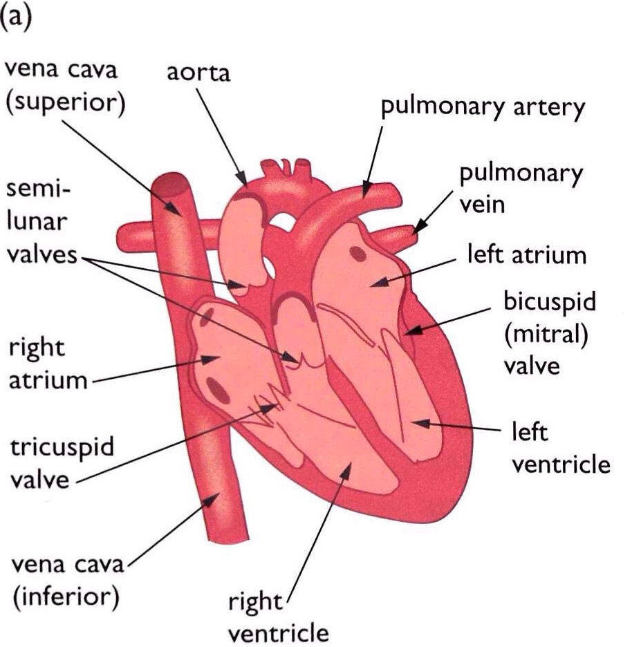
Label the heart — Science Learning Hub In this interactive, you can label parts of the human heart. Drag and drop the text labels onto the boxes next to the diagram. Selecting or hovering over a box will highlight each area in the diagram. Right ventricle Right atrium Left atrium Pulmonary artery Left ventricle Pulmonary vein Semilunar valve Vena cava Aorta Download Exercise Tweet

External structure of the heart with labels
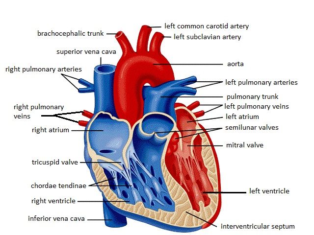
Correctly Label The Following External Anatomy Of The Anterior Heart ... The external anatomy of the human heart consists of the four chambers that form the apex of the heart. Each chamber has an apex that corresponds to a box. There are two boxes on each side of the heart: the atria and the ventricles. The left atrium is a branching organ. The pulmonary trunk contains the aorta and pulmonary veins. Human Heart Diagram Labeled | Science Trends The endocardium is the inner portion of the outer wall, and the endocardium is what contacts the blood in the heart. The heart's atrioventricular valves are structures that join the atria and ventricles of the heart together. This group of valves is comprised of the tricuspid valve and the mitral valve. › physiology-of-the-heartPhysiology of the Heart | Boundless Anatomy and Physiology ... The first wave on an ECG is the P wave, indicating atrial depolarization in which the atria contract (atrial systole ). The P wave is the first wave on the ECG because the action potential for the heart is generated in the sinoatrial (SA) node, located on the atria, which sends action potentials directly through Bachmann's bundle to depolarize the atrial muscle cells.
External structure of the heart with labels. Structure of the Heart | SEER Training The human heart is a four-chambered muscular organ, shaped and sized roughly like a man's closed fist with two-thirds of the mass to the left of midline. The heart is enclosed in a pericardial sac that is lined with the parietal layers of a serous membrane. The visceral layer of the serous membrane forms the epicardium. Layers of the Heart Wall en.wikipedia.org › wiki › DapagliflozinDapagliflozin - Wikipedia Medical uses. Dapagliflozin is used along with diet, exercise and usually with other glucose lowering medications, to improve glycaemic control in adults with type 2 diabetes and to reduce the risk of hospitalisation for heart failure among adults with type 2 diabetes and known cardiovascular disease or other cardiovascular risk factors (including high blood pressure, high cholesterol and ... Heart Diagram with Labels and Detailed Explanation - BYJUS Diagram of Heart. The human heart is the most crucial organ of the human body. It pumps blood from the heart to different parts of the body and back to the heart. The most common heart attack symptoms or warning signs are chest pain, breathlessness, nausea, sweating etc. The diagram of heart is beneficial for Class 10 and 12 and is frequently ... external structure of heart How would you label the structures (both external and internal) of a we have 8 Pictures about How would you label the structures (both external and internal) of a like How would you label the structures (both external and internal) of a, Blood Vessels Flashcards | Easy Notecards and also Toad Dissection (how to/how not to) - YouTube.
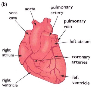
Ch. 19 Circulatory System- heart Flashcards - Quizlet Correctly label the external anatomy of the anterior heart. Place the labels in order denoting the flow of blood through the pulmonary circuit beginning with the right atrium and ending in the left atrioventricular valve. The first and last structures are given. Right atrium 1. tricuspid valve 2. right ventricle 3. pulmonary valve en.wikipedia.org › wiki › The_TenorsThe Tenors - Wikipedia The Tenors (formerly known as The Canadian Tenors) are a vocal group consisting of Victor Micallef, Fraser Walters, and Clifton Murray.They perform operatic pop music that is a mixture of classical and pop, featuring songs such as "The Prayer", Panis angelicus, and Leonard Cohen's Hallelujah. › programs › heart-weekHeart Week 2022 | The Heart Foundation Heart Health Checks present a valuable opportunity for healthcare professionals to engage with their patients about their risk of developing cardiovascular disease and ways to lower this risk. pmt.physicsandmathstutor.com › download › BiologyPractical notes - SP 2.3c Dissection of a Mammalian Heart ... The mammalian heart is a muscular pump that pushes blood around the body. It consists of four chambers and associated blood vessels . The left and right side of the heart is separated by a muscular wall, the septum . Recall the structure of the heart in the diagram below:
Heart anatomy: Structure, valves, coronary vessels | Kenhub The heart is shaped as a quadrangular pyramid, and orientated as if the pyramid has fallen onto one of its sides so that its base faces the posterior thoracic wall, and its apex is pointed toward the anterior thoracic wall. The Anatomy of the Heart, Its Structures, and Functions The heart is the organ that helps supply blood and oxygen to all parts of the body. It is divided by a partition (or septum) into two halves. The halves are, in turn, divided into four chambers. The heart is situated within the chest cavity and surrounded by a fluid-filled sac called the pericardium. This amazing muscle produces electrical ... hbr.org › 2009 › 09How Strategy Shapes Structure - Harvard Business Review See Industrial Market Structure and Economic Performance, F. M. Sherer (Chicago: Rand McNally, 1970). 2. See Blue Ocean Strategy , W. Chan Kim and Renée Mauborgne (Harvard Business Press, 2005). Layers of the heart: Epicardium, myocardium, endocardium - Kenhub The myocardium is functionally the main constituent of the heart and the thickest layer of all three heart layers. It is a muscle layer that enables heart contractions. Histologically, the myocardium is comprised of cardiomyocytes.Cardiomyocytes have a single nucleus in the center of the cell, which helps to distinguish them from skeletal muscle cells that have multiple nuclei dispersed in the ...
Heart Anatomy Using Models
external structure human heart diagram Drawing of the brain with labels. Labels brain drawing ear anatomy functions human diagram labeled clipartmag. Sheep dissection anatomy heart side dissected pig human class physiology card labeled valve science cow septum blood term cardiac interventricular
iGCSE Biology - Gross Structure Of The Heart - BioChem Tuition
External anterior heart labeling Quiz - PurposeGames.com An unregistered player played the game 5 hours ago About this Quiz This is an online quiz called External anterior heart labeling There is a printable worksheet available for download here so you can take the quiz with pen and paper. Total Points 0 Get started! Today's Rank -- 0 Today 's Points Points 27 to score the 27 points available

Heart structure and function — structure and function of the heart cardiac output
Heart Anatomy: Heart Dissection - University of Washington The major vessels of the heart are found at the base of the heart, along with the upper chambers, the right atrium (C) and left atrium (D). The atria are collapsed, but in a functioning heart, they would be stretched full of blood. The majority of the heart tissue consists of the ventricles. The left ventricle (F) is stiff and solid because it ...

Science&Life: Heart Dissection
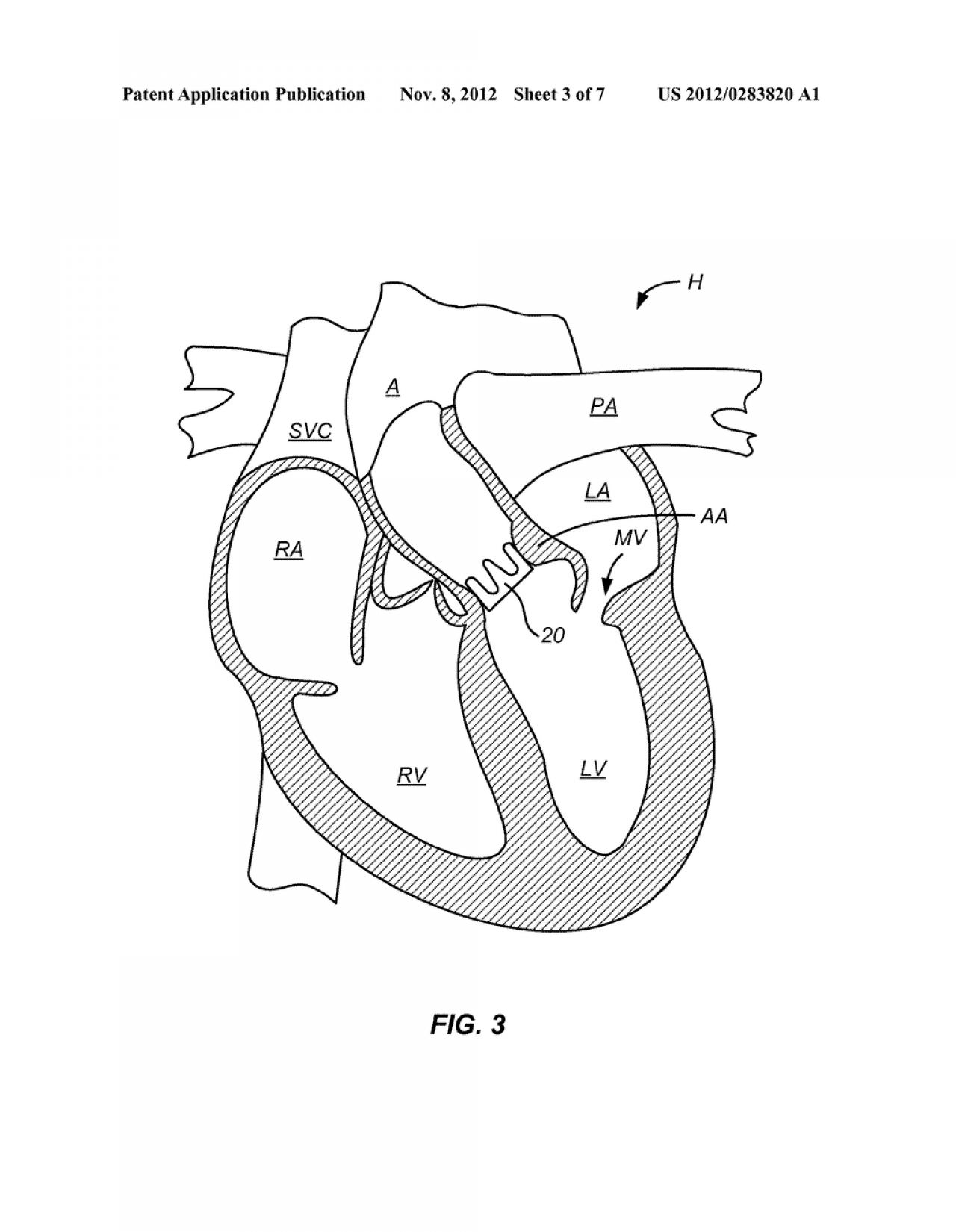
Solved Help Label the external anatomy on this posterior - Chegg Question: Help Label the external anatomy on this posterior view of a mammalian heart by clicking and dragging the labels to the correct location Coronary sinus Apex of heart Lert atrium Posterior interventricular branch of LCA Left pulmonary artery Left ventricle Left pulmonary veins Aortic arch This problem has been solved! See the answer

Biology Diagrams,Images,Pictures of Human anatomy and physiology : Heart External Structure ...
Correctly Label The Following Internal Anatomy Of The Heart The aorta, or aortic arch, is the outermost layer of the heart. The left ventricle is covered with the ventricular aorta, and the pulmonary veins are located inside the aorta. The two atria, the left and right aorta, and the right aortic arch are all external organs. These organs carry oxygen-rich blood to the body.
iGCSE Biology - Gross Structure Of The Heart - BioChem Tuition
Human Heart (Anatomy): Diagram, Function, Chambers, Location in Body Electrocardiogram (ECG or EKG): A tracing of the heart's electrical activity. Electrocardiograms can help diagnose many heart conditions. Echocardiogram: An ultrasound of the heart. An ...
Free Blank Heart Diagram, Download Free Blank Heart Diagram png images, Free ClipArts on Clipart ...
Label the Heart - The Biology Corner Shows a picture of a heart with letters and blanks for practice with labeling the parts of the heart and tracing the flow of blood within the heart.

Human Anatomy - Beverly Afzali
Heart - External Features - Anatomy QA Location of heart: Heart lies in the middle mediastinum. 1/3rd of the heart lies to the right and 2/3rd to the left of the midline. It lies opposite to T5 - T8 vertebrae in supine position & T6 - T9 vertebrae in erect position. Dimensions of heart: Base to apex-12cm; Transversely- 8-9cm; Anteroposteriorly- 6cm.
/heart_exterior_anatomy-577d5cc23df78cb62c942f06.jpg)
Anatomy of the Heart - Diagram View
Chapter 22 Heart Flashcards - Quizlet Label the coronary arteries in an anterior view of the heart. Label the order that blood flows through in the heart, using the arrows as guides. Label the components of the heart wall. Label the components of the heart as seen from a posterior view. Label the major coronary veins. Label the components of the conduction system.

Biology 156
A Labeled Diagram of the Human Heart You Really Need to See The human heart, comprises four chambers: right atrium, left atrium, right ventricle and left ventricle. The two upper chambers are called the left and the right atria, and the two lower chambers are known as the left and the right ventricles. The two atria and ventricles are separated from each other by a muscle wall called 'septum'.
Unlabelled Diagram Of The Heart - Cliparts.co
How to Draw the Internal Structure of the Heart (with Pictures) To draw the internal structure of a human heart, follow the steps below. Part 1 Finding a Diagram 1 To find a good diagram, go to Google Images, and type in "The Internal Structure of the Human Heart". Find an image that displays the entire heart, and click on it to enlarge it. 2 Find a piece of paper and something to draw with.

u414adad: heart diagram without labels
Solved Art-Labeling Activity: Overview of the external - Chegg art-labeling activity: overview of the external anatomy of the heart anterior view res great cardiac vein aortic arch right coronary artery left coronary artery left pulmonary veins ascending aorta left pulmonary artery anterior interventricular artery superior vena cava pulmonary trunk auricle of left atrium circumflex artery auricle of right …

How Your Heart Works | Health Resources | Wellness Library | UPMC Pinnacle
The structure of the heart - Structure and function of the heart ... Each side of the heart consists of an atrium and a ventricle which are two connected chambers. The atria (plural of atrium) are where the blood collects when it enters the heart. The ventricles...

Biology Diagrams,Images,Pictures of Human anatomy and physiology : Heart External Structure ...
Heart Anatomy: Labeled Diagram, Structures, Function, and Blood Flow Let's begin with the chambers of the heart. There are 4 chambers, labeled 1-4 on the diagram below. To help simplify things, we can convert the heart into a square. We will then divide that square into 4 different boxes which will represent the 4 chambers of the heart.
Heart. Structure of the Heart. Divisions of the Heart
Heart Anatomy Labeling Game - PurposeGames.com This is an online quiz called Heart Anatomy Labeling Game. There is a printable worksheet available for download here so you can take the quiz with pen and paper. Your Skills & Rank. Total Points. 0. Get started! Today's Rank--0. Today 's Points. One of us! Game Points. 19. You need to get 100% to score the 19 points available.

1000+ images about School on Pinterest | Heart diagram, Physician assistant and Medical
Human Heart - Diagram and Anatomy of the Heart - Innerbody Because the heart points to the left, about 2/3 of the heart's mass is found on the left side of the body and the other 1/3 is on the right. Anatomy of the Heart Pericardium. The heart sits within a fluid-filled cavity called the pericardial cavity. The walls and lining of the pericardial cavity are a special membrane known as the pericardium.
Post a Comment for "40 external structure of the heart with labels"