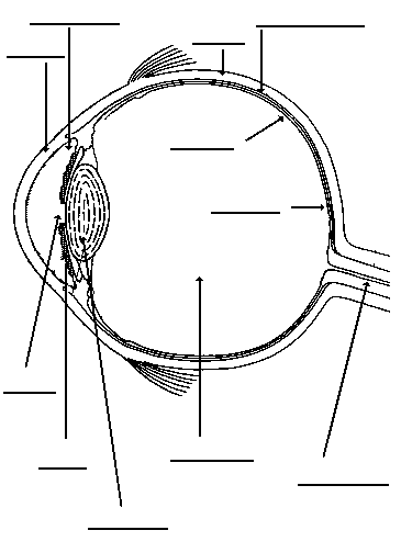
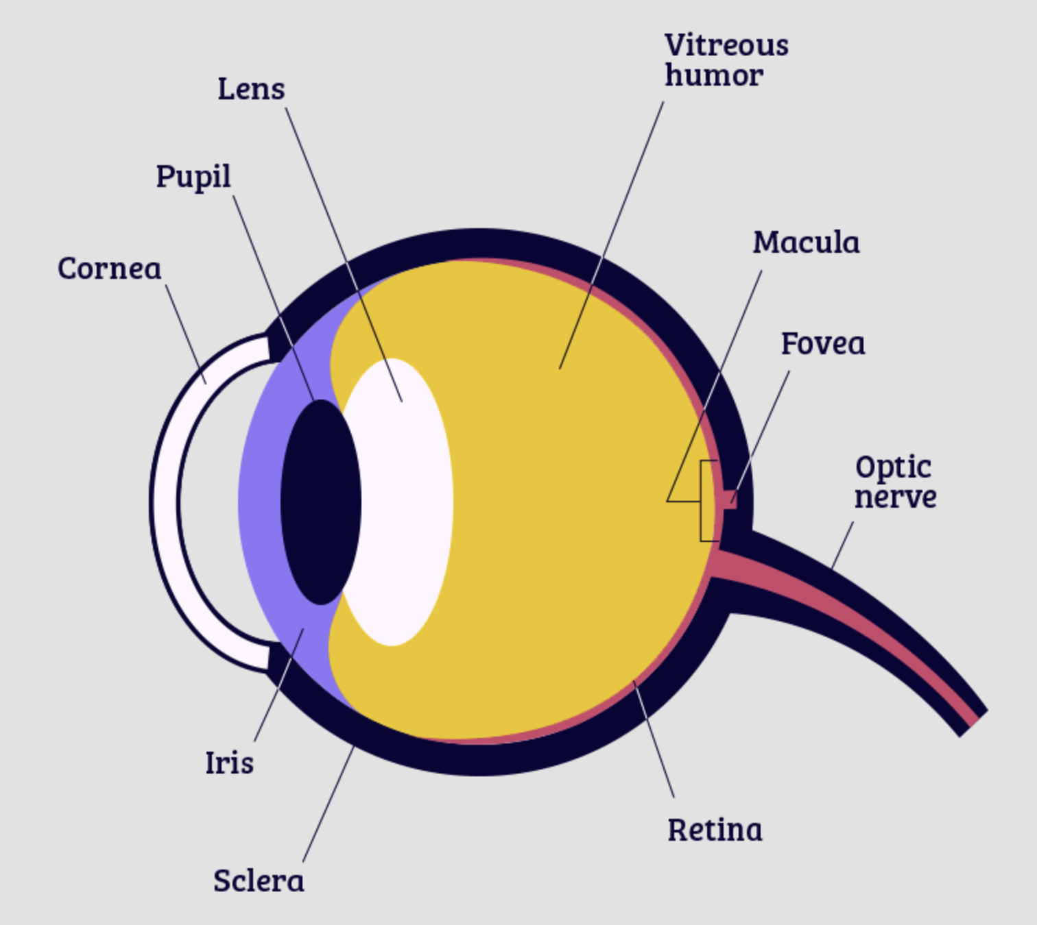
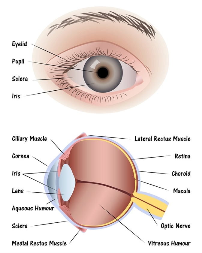
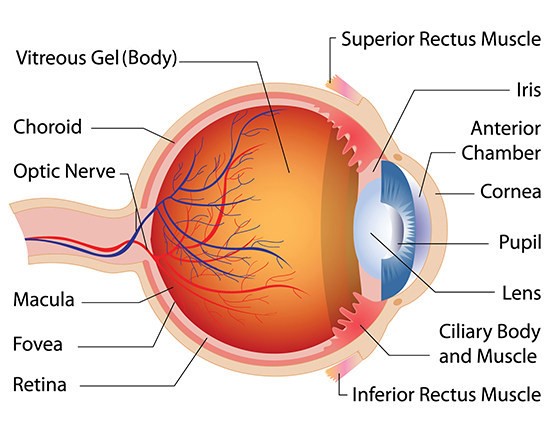
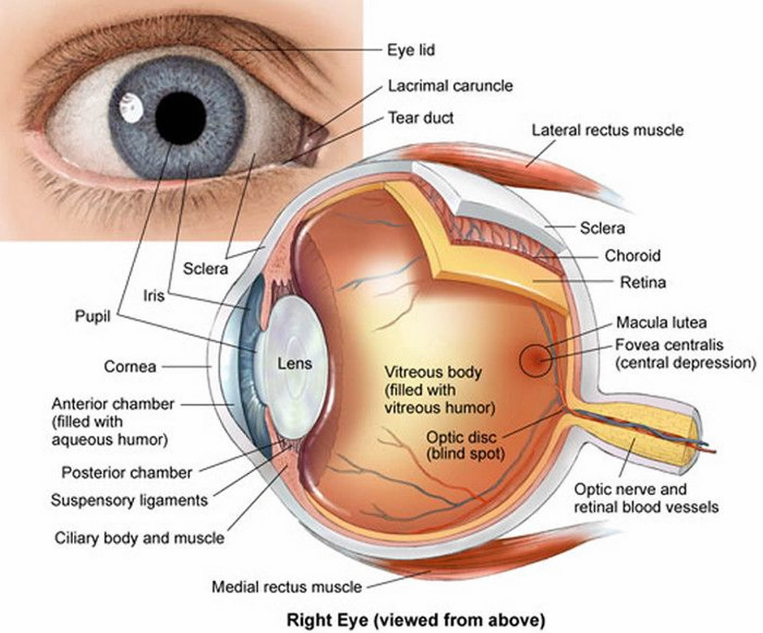
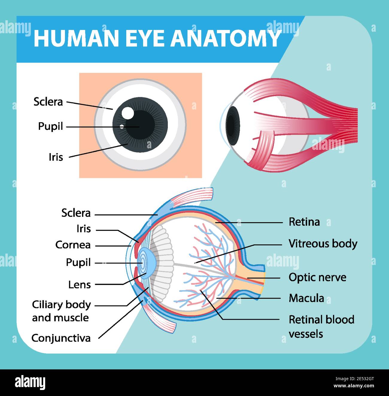
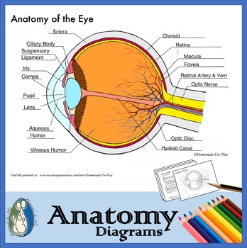
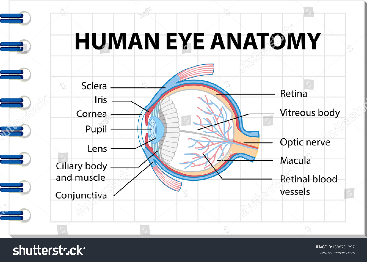
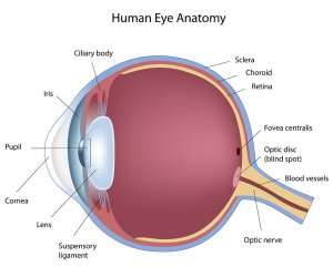
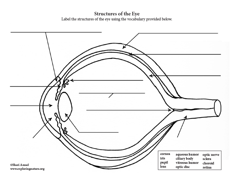
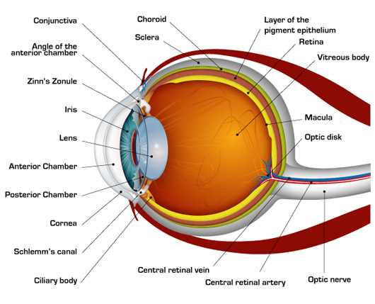
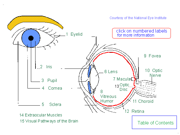
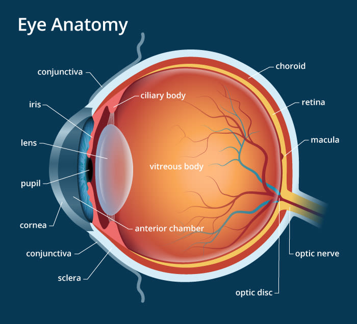
45 eye diagram and labels
Parts of the Microscope with Labeling (also Free Printouts) It is used to observe things that cannot be seen by the naked eye. Table of Contents 1. Eyepiece 2. Body tube/Head 3. Turret/Nose piece 4. Objective lenses 5. Knobs (fine and coarse) 6. Stage and stage clips 7. Aperture 9. Condenser 10. Condenser focus knob 11. Iris diaphragm 12. Diopter adjustment 13. Arm 14. Specimen/slide 15. Labeled imaging anatomy cases | Radiology Reference Article ... This article lists a series of labeled imaging anatomy cases by body region and modality. Brain CT head: non-contrast axial CT head: non-contrast coronal CT head: non-contrast sagittal CT head: angiogram axial CT head: angiogram coronal CT...
Simple Microscope - Diagram (Parts labelled), Principle, Formula and Uses The working principle of a simple microscope is that when a lens is held close to the eye, a virtual, magnified and erect image of a specimen is formed at the least possible distance from which a human eye can discern objects clearly. Magnification formula The magnification power of a simple microscope is expressed as: M = 1 + D/F Where

Eye diagram and labels
Eye Diagram Quiz - ProProfs Quiz Try this amazing Eye Diagram Quiz quiz which has been attempted 5204 times by avid quiz takers. Also explore over 78 similar quizzes in this category. Take Quizzes. Animal; Nutrition; ... Quiz: Label The Parts Of The Eye. People say that the eyes are the windows to a person's soul. In the class today, we covered parts of the eye, and what ... Can Eye Drops Improve Vision in Myopia? - All About Vision Atropine eye drops have been used in doctor's offices for many years. They dilate the pupil and paralyze the accommodative system of the eye. Eye doctors usually use 1% atropine eye drops in their offices for pupil dilation. For myopia control, children require a lower dose such as 0.01%, 0.025% or 0.05% concentration. Microscope Types (with labeled diagrams) and Functions The working principle of a simple microscope is that when a lens is held close to the eye, a virtual, magnified and erect image of a specimen is formed at the least possible distance from which a human eye can discern objects clearly. Simple microscope labeled diagram Simple microscope functions It is used in industrial applications like:
Eye diagram and labels. Parts of a microscope with functions and labeled diagram - Microbe Notes The coarse adjustment knob moves the stage up and down to bring the specimen into focus. The fine adjustment knob brings the specimen into sharp focus under low power and is used for all focusing when using high-power lenses. Q. List down the 18 parts of a Microscope. 1. Ocular Lens (Eye Piece) 2. Diopter Adjustment 3. Head 4. Nose Piece 5. The Lens: Anatomy, Function, and Treatment - Verywell Health Tests. The lens is a curved structure in the eye that that bends light and focuses it for the retina to help you see images clearly. The crystalline lens, a clear disk behind the iris, is flexible and changes shape to help you see objects at varying distances. As you age, the lens may become weaker or damaged. Description of 18 Structures in Labeled Diagram of the Eye VI Student's Audio Description of Eye Structures on a Diagram Anatomy of the Eye - Verywell Health Cornea. Pupil. Iris. Crystalline Lens. Aqueous Humor. The human eye is an organ that detects light and sends signals along the optic nerve to the brain. Perhaps one of the most complex organs of the body, the eye is made up of several parts—and each individual part contributes to your ability to see.
Eye Anatomy: 16 Parts of the Eye & Their Functions - Vision Center The following are parts of the human eyes and their functions: 1. Conjunctiva The conjunctiva is the membrane covering the sclera (white portion of your eye). The conjunctiva also covers the interior of your eyelids. Conjunctivitis, often known as pink eye, occurs when this thin membrane becomes inflamed or swollen. Quiz: Label The Parts Of The Eye - ProProfs Quiz Quiz: Label The Parts Of The Eye. Do you know the anatomy of the human eye very well? Can you label the parts of the eye in the quiz below? Give it a try and evaluate yourself. The eye has many important parts, each with different functions, including the cornea, pupil, sclera, and many more. Can you tell where these parts are located and what ... What Eye Problems Look Like - WebMD Cover one eye and stare at the center dot in this Amsler grid from a distance of 12 to 15 inches. (You can wear your reading glasses.) Do you see wavy, broken, or blurry lines? Are any areas warped... External and internal eye anatomy - MedlinePlus Overview. The cornea allows light to enter the eye. As light passes through the eye the iris changes shape by expanding and letting more light through or constricting and letting less light through to change pupil size. The lens then changes shape to allow the accurate focusing of light on the retina. Light excites photoreceptors that ...
Eye Anatomy and Physiology a Complete Detail - Study Read The anatomy and physiology of the eye are highly organized and effective despite being small. Eye Anatomy and Physiology Eyes are spheroid shape organs fitted into the two orbitals of the skull. There are three major parts in each eye like The sclera (fibrous layer) Choroid layer Retina Eyes diagram showing the entire structure The sclera How To Make Eye-Catching and Professional Diagrams With Visio To make a simple diagram, you can use the includes shapes and symbols. Just type in labels and link your symbols with the connector tool. By selecting different color and style options from the ribbon menu, you can create an eye-catching and professional diagram in a matter of minutes. New Features That Enable Great Diagrams anatomy and physiology of eye pdf free download Labeled Eye Diagram Human Eye Diagram Eye Anatomy Diagram Of The Eye The development of a beak has led to evolution of a specially.. Anatomy And Physiology of Human body. Learning Resources Inc 2008 Other. Anatomy and Physiology 2e is developed to meet the scope and sequence for a two-semester human anatomy and physiology course for life ... Chemical Eye Burns: When Should I Get Medical Help? - All About Vision Read labels on chemicals. Learn how to use the chemical properly and what to do if it gets in your eyes. When possible, swap out more dangerous chemicals like ammonia for milder cleaners. Stay safe from fumes. It's not just physical contact that can harm the eyes. Fumes can cause damage to the mucous membranes of the eyes.
Anatomy and Structure of the Human Eye (With Diagrams) In the diagram, the artery is shown in red, and the vein is in blue. Tear Duct This is a small tube running from the eye to the nasal cavity. Tears drain from the eyes into the nose through the tear duct. This is why teary eyes are usually accompanied by a runny nose. Schematic animation of the human eyeball
Parts Of The Eye Labeled Diagram Model And Their - SUNGLASSKY Parts of the eye-labeled diagram model are divided into three groups: the external outer layer, the middle layer, and the inner back layer. The outer layer is responsible for protecting the eye from environmental toxins and debris. The middle layer includes cells that allow light to enter and travel through the back layer to the retina.
WHMIS 2015 - Pictograms : OSH Answers - Canadian Centre for ... The exclamation mark pictogram is used for the following classes and categories: Acute toxicity - Oral, Dermal, Inhalation (Category 4) Skin corrosion/irritation - Skin irritation (Category 2) Serious eye damage/eye irritation - Eye irritation (Category 2 and 2A) Respiratory or skin sensitization - Skin sensitizer (Category 1, 1A and 1B)
Microscope, Microscope Parts, Labeled Diagram, and Functions The Iris Diaphragm is located above the condenser lens and below the microscope stage. The different sized holes in the diaphragm helps to vary the size of the cone and intensity of light that is projected upward into the slide. However, there is no set rule regarding which setting to use for a particular power.
Anatomy of the Eye | BrightFocus Foundation Cornea: The outer, transparent structure at the front of the eye that covers the iris, pupil and anterior chamber; it is the eye's primary light-focusing structure. Drusen: Deposits of yellowish extra cellular waste products that accumulate within and beneath the retinal pigmented epithelium (RPE) layer. Fovea: The pit or depression at the ...
Parts of Human Eye and Their Functions | MD-Health.com The iris is the area of the eye that contains the pigment which gives the eye its color. This area surrounds the pupil, and uses the dilator pupillae muscles to widen or close the pupil. This allows the eye to take in more or less light depending on how bright it is around you. If it is too bright, the iris will shrink the pupil so that they ...
Anatomy of the eye: Quizzes and diagrams | Kenhub One of our favorite ways to get to grips with all of the parts of the eye is by utilizing labeled diagrams. On a diagram of the eye, we can see all of the relevant structures together on one image. This helps us to understand how each one is situated and related to the other. Labeled diagram of the eye
20/20 Vision - Activity - TeachEngineering Require the diagram to include a light source, an object to be seen, and the eye viewing the object. In the diagram, have them label the cornea, iris and pupil, lens, and retina. Additionally, challenge students to add a second lens (glasses) to the diagram and describe the effect on vision of the added lens. Troubleshooting Tips
Eye Anatomy | Blood supply - Orbit - Extraocular muscles - Geeky Medics In total, there are seven extraocular muscles. Six of these are responsible for the movement of the eye, with the seventh being responsible for the movement of the superior eyelid. The muscles responsible for the movement of the eye may be divided into the four recti muscles and the two oblique muscles. Table 1.
Eye anatomy: Muscles, arteries, nerves and lacrimal gland - Kenhub The wall of the eyeball is three-layered; with the sclera as the outer layer (continuous with the cornea), choroid as the middle vascular layer (continuous with the ciliary body and iris ), and the retina as the innermost layer. You can study the anatomy of the eyeball in detail through this study unit. Anatomy of the eyeball Explore study unit
Microscope Parts, Function, & Labeled Diagram - slidingmotion Microscope parts labeled diagram gives us all the information about its parts and their position in the microscope. Microscope Parts Labeled Diagram The principle of the Microscope gives you an exact reason to use it. It works on the 3 principles. Magnification Resolving Power Numerical Aperture. Parts of Microscope Head Base Arm Eyepiece Lens
Microscope Types (with labeled diagrams) and Functions The working principle of a simple microscope is that when a lens is held close to the eye, a virtual, magnified and erect image of a specimen is formed at the least possible distance from which a human eye can discern objects clearly. Simple microscope labeled diagram Simple microscope functions It is used in industrial applications like:
Can Eye Drops Improve Vision in Myopia? - All About Vision Atropine eye drops have been used in doctor's offices for many years. They dilate the pupil and paralyze the accommodative system of the eye. Eye doctors usually use 1% atropine eye drops in their offices for pupil dilation. For myopia control, children require a lower dose such as 0.01%, 0.025% or 0.05% concentration.
Eye Diagram Quiz - ProProfs Quiz Try this amazing Eye Diagram Quiz quiz which has been attempted 5204 times by avid quiz takers. Also explore over 78 similar quizzes in this category. Take Quizzes. Animal; Nutrition; ... Quiz: Label The Parts Of The Eye. People say that the eyes are the windows to a person's soul. In the class today, we covered parts of the eye, and what ...










/GettyImages-695204442-b9320f82932c49bcac765167b95f4af6.jpg)
:background_color(FFFFFF):format(jpeg)/images/library/11201/overview_parts_of_the_eye_labelled_diagram.jpg)


/GettyImages-695204442-b9320f82932c49bcac765167b95f4af6.jpg)


Post a Comment for "45 eye diagram and labels"