45 cell wall diagram with labels
Intestinal transgene delivery with native E. coli chassis allows ... 4.8.2022 · Native E. coli strains isolated from mouse stool are genetically engineered for long-term engraftment in the conventional mouse gut and enable long-term systemic effects on the host, such as improvements in insulin sensitivity in mouse models of type 2 diabetes. Cell Organelles- Definition, Structure, Functions, Diagram - Microbe Notes In a plant cell, the cell wall is made up of cellulose, hemicellulose, and proteins while in a fungal cell, it is composed of chitin. A cell wall is multilayered with a middle lamina, a primary cell wall, and a secondary cell wall. The middle lamina contains polysaccharides that provide adhesion and allow binding of the cells to one another.
03 Label the Cell Diagram | Quizlet Start studying 03 Label the Cell. Learn vocabulary, terms, and more with flashcards, games, and other study tools.
Cell wall diagram with labels
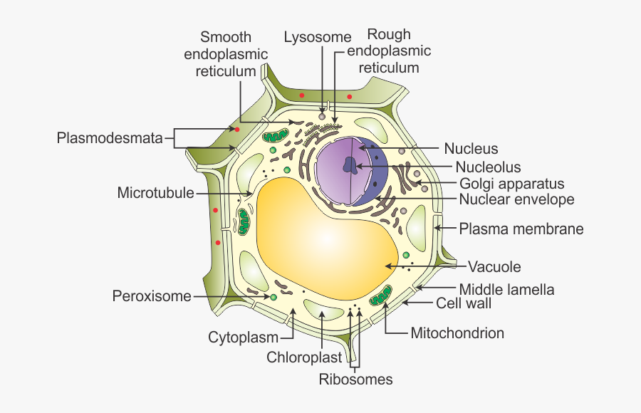
Draw a neat diagram of spirogyra and label the cell wall ? - QnA ... Draw a neat diagram of spirogyra and label the cell wall ? - QnA Explained Labeled Plant Cell With Diagrams | Science Trends The parts of a plant cell include the cell wall, the cell membrane, the cytoskeleton or cytoplasm, the nucleus, the Golgi body, the mitochondria, the peroxisome's, the vacuoles, ribosomes, and the endoplasmic reticulum. Parts Of A Plant Cell The Cell Wall Let's start from the outside and work our way inwards. Bacteria in Microbiology - shapes, structure and diagram - Jotscroll The bacteria shapes, structure, and labeled diagrams are discussed below. Table of Contents [ show] Sizes The sizes of bacteria cells that can infect human beings range from 0.1 to 10 micrometers. Some larger types of bacteria such as the rickettsias, mycoplasmas, and chlamydias have similar sizes as the largest types of viruses, the poxviruses.
Cell wall diagram with labels. Elodea Leaf Cell Diagram Elodea Leaf Cell Diagram The Elodea leaf is composed of two layers of cells. Only one layer of cells is in focus when using the high. Examining elodea (pondweed) under a compound microscope. solution) and a coverslip and observe the chloroplasts (green structures) and the cell walls. How to Create 3D Plant Cell and Animal Cell Models for 25.8.2022 · Cell Wall: Found only in plant cells, the cell wall surrounds the cell membrane. The cell wall is stiff and rigid, and it provides additional protection and support to the cell. Chloroplasts: Found only in plant cells, chloroplasts produce food (energy) for the cell by converting sunlight, carbon dioxide, and water into sugars. Cell: Structure and Functions (With Diagram) - Biology Discussion Eukaryotic Cells: 1. Eukaryotes are sophisticated cells with a well defined nucleus and cell organelles. 2. The cells are comparatively larger in size (10-100 μm). 3. Unicellular to multicellular in nature and evolved ~1 billion years ago. 4. The cell membrane is semipermeable and flexible. 5. These cells reproduce both asexually and sexually. Human Cell Diagram, Parts, Pictures, Structure and Functions Diagram of the human cell illustrating the different parts of the cell. Cell Membrane. The cell membrane is the outer coating of the cell and contains the cytoplasm, substances within it and the organelle. It is a double-layered membrane composed of proteins and lipids. The lipid molecules on the outer and inner part (lipid bilayer) allow it to ...
Nervous System – Medical Terminology for Healthcare Professions Introduction to the Nervous System. The picture you have in your mind of the nervous system probably includes the brain, the nervous tissue contained within the cranium, and the spinal cord, the extension of nervous tissue within the vertebral column.That suggests it is made of two organs—and you may not even think of the spinal cord as an organ—but the nervous system … TIMWOOD [Definition of the 7 Wastes] | Creative Safety Supply It's easy to remember the 7 Wastes of Lean with the acronym TIMWOOD. Learn what each letter stands for and how to counteract each waste. TIMWOOD stands for the Seven Wastes of Lean: transportation, inventory, motion, waiting, overproduction, over-processing, and defects. Plant Cells: Labelled Diagram, Definitions, and Structure - Research Tweet The cell wall is made of cellulose and lignin, which are strong and tough compounds. Plant Cells Labelled Plastids and Chloroplasts Plants make their own food through photosynthesis. Plant cells have plastids, which animal cells don't. Plastids are organelles used to make and store needed compounds. Chloroplasts are the most important of plastids. Plant Cell Wall- Definition, Structure, Functions, Diagram Structure of Plant Cell wall It is derived from the living protoplast. It consists of the middle lamella, primary cell wall, plasmodesmata, secondary cell wall, and pits. Middle lamella After the cytokinesis, it is the first-formed layer. It is present in between the two adjacent cells. It is made up of calcium and magnesium pectate.
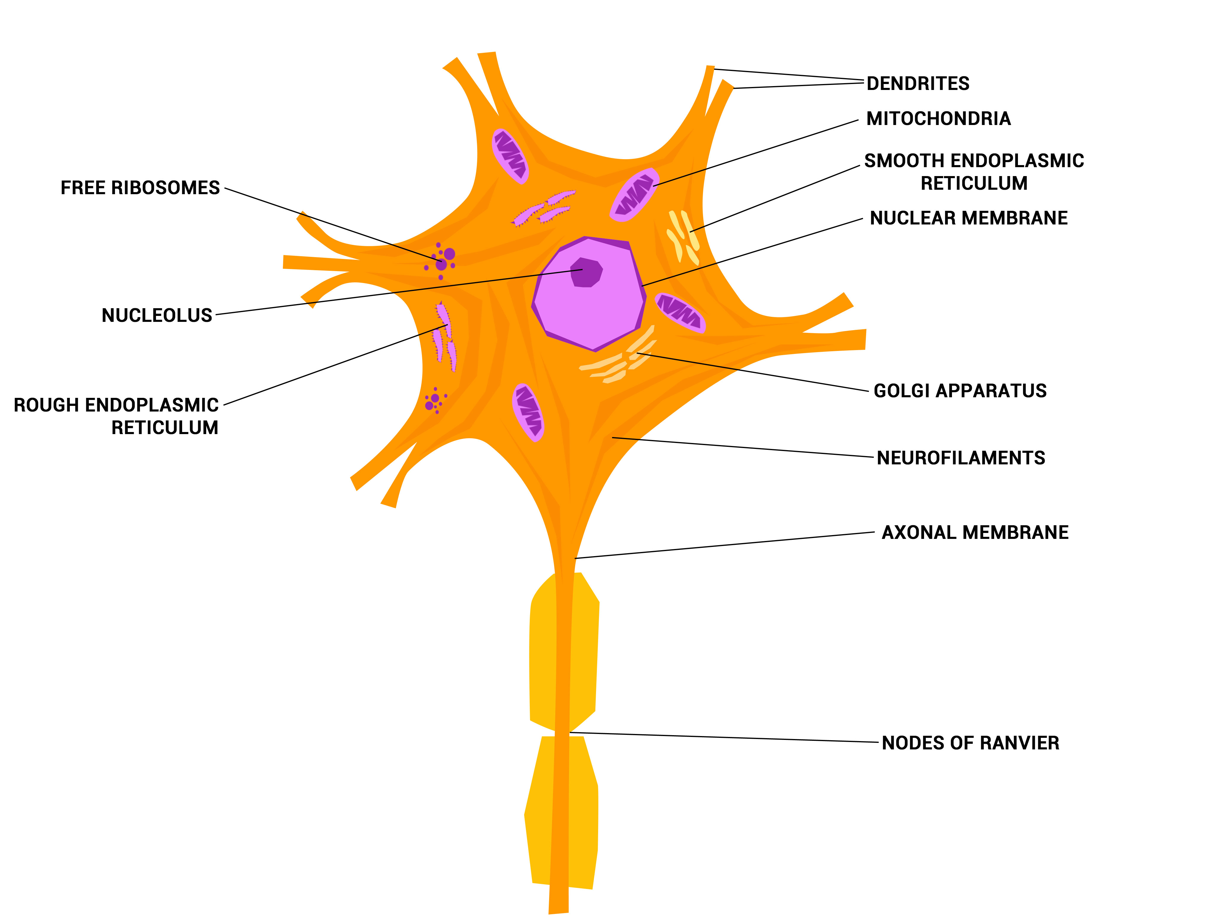
Cell Wall Function, Structure and Diagram - jotscroll.com It helps to control the expansion of cells during water intake. Cell walls function in the control of cell expansion. The elastic (reversible) and plastic (irreversible) nature of the wall is a major factor in the growth of plant cells. When water moves into the cell, it causes the protoplast to press against the wall. Free Cell Diagram Software with Free Templates - EdrawMax - Edrawsoft A plant cell diagram has a cell wall, cell membrane, nucleus, organelles, endoplasmic reticulum, ribosome, plastids, mitochondria, and multiple vesicles. Animal Cell Diagram An animal cell diagram describes a cell structure enclosed by a plasma member, and it has a nucleus with a membrane and organelles. Neuron Diagram Animal Cells: Labelled Diagram, Definitions, and Structure - Research Tweet Cell Organelles Plant Cells: Animal Cells: Cell wall: Present (made up of cellulose) Absent: Shape: Rectangular (fixed shape) Round (irregular shape) Vacuole: One, large central vacuole taking up to 90% of cell volume. One or more small vacuoles (much smaller than plant cells). Centrioles: Only present in lower plant forms (e.g. chlamydomonas) An Overview of Hyphae in Fungi, Their Function and Types. 10.1.2022 · Fungi have their cell wall made up of chitin. Their body is composed of long thread-like filaments or tubes known as hyphae . In singular form, this structure is referred to as a hypha.
PDF Human Cell Diagram, Parts, Pictures, Structure and Functions Human Cell Diagram, Parts, Pictures, Structure and Functions The cell is the basic functional in a human meaning that it is a self-contained and fully operational living entity. Humans are multicellular organisms with various different types of cells that work together to sustain life. Other non-cellular components in the body include water ...

parts of a cell .pdf - Organelle Function(Job Cell wall The cell wall surrounds the plasma ...
American Express Printable colored labels. Each of our true color materials are both laser and inkjet printable and corresponds to colors in the Pantone Matching System (PMS). Pantone values for our true color label collection: Light Tan (TC) - PMS 726 U True Blue (TB) - PMS 2925 U True Gray (TE) - PMS 421 U True Green (TG) - PMS 361 U True Purple (TP) - PMS 2645 U. Akhal-Teke horses are …
Animal Cell Labelling Activity | Primary Resources | Twinkl A colorful resource which covers the parts of an animal cell including the nucleus, cell wall, cytoplasm, and mitochondria. Lower, middle and higher ability versions are available. ... this Plant Cell Diagram is a similar labelling activity for plant cells. ... you could try using this Polar Bear Animal Diagram with Labels.
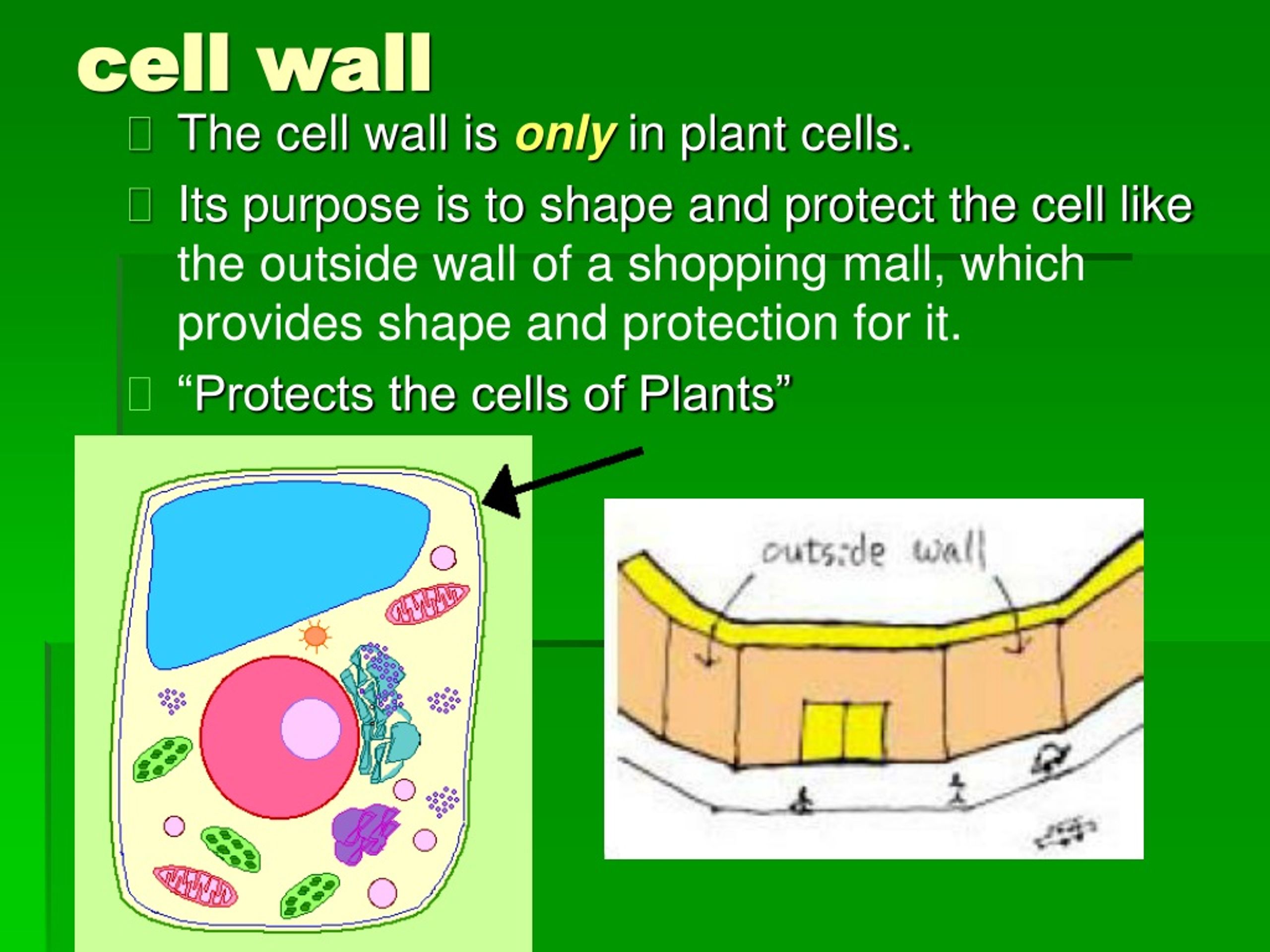
Cell Wall - Definition, Cell Wall Function, Cell Wall Layers - BYJUS The plant cell wall is generally arranged in 3 layers and composed of carbohydrates, like pectin, cellulose, hemicellulose and other smaller amounts of minerals, which form a network along with structural proteins to form the cell wall. The three major layers are: Primary Cell Wall The Middle Lamella The Secondary Cell Wall
Label Cell Parts | Plant & Animal Cell Activity | StoryboardThat Click "Start Assignment". Find diagrams of a plant and an animal cell in the Science tab. Using arrows and Textables, label each part of the cell and describe its function. Color the text boxes to group them into organelles found in only animal cells, organelles found in only plant cells, and organelles found in both cell types.
A Labeled Diagram of the Plant Cell and Functions of its Organelles A Labeled Diagram of the Plant Cell and Functions of its Organelles We are aware that all life stems from a single cell, and that the cell is the most basic unit of all living organisms. The cell being the smallest unit of life, is akin to a tiny room which houses several organs. Here, let's study the plant cell in detail...
Label the cell - Teaching resources - Wordwall Label Plant and Animal Cell Labelled diagram by Eawilson the cell Match up by Elenagp9149 5.6 Label the sentence Labelled diagram by Christianjolene Label the Electromagnetic Spectrum Labelled diagram by Elizabetheck G6 G7 G8 Science 5.7 Label the sentence Labelled diagram by Christianjolene The cell Anagram by Thepowerhouse G7 G8 Science
Cell Wall: Definition, Structure & Function (with Diagram) In general, fungi with cell walls have three layers: chitin, glucans and proteins. As the innermost layer, chitin is fibrous and made up of polysaccharides. It helps make the fungi cell walls rigid and strong. Next, there is a layer of glucans, which are glucose polymers, crosslinking with chitin.
History of cell membrane theory - Wikipedia Cell theory has its origins in seventeenth century microscopy observations, but it was nearly two hundred years before a complete cell membrane theory was developed to explain what separates cells from the outside world. By the 19th century it was accepted that some form of semi-permeable barrier must exist around a cell. Studies of the action of anesthetic molecules led to …

In The Figure Which Diagram Of A Cell Wall Has A Structure That Protects Against Osmotic Lysis ...
Structure of Cell Wall (With Diagram) | Plants - Biology Discussion Cells with secondary wall consist of five layers a three layered secondary wall, the primary wall and the middle lamella. In some cells, such as primary xylem, the secondary thickening materials are laid down in such a way that various patterns are formed on the cell wall, e.g. annular, spiral, reticulate, scalariform and pitted.
Cell wall structure with plant cellular parts description outline diagram Cell wall structure with plant cellular parts description outline diagram. Illustration about physiology, structure, vector, educational, molecular, leaf, parts, biomass - 225074534 ... Root hair cell collecting mineral nutrients and water from soil, biological labeled plant system diagram. Gram positive versus negative cell wall structure ...
Label animal cell - Teaching resources - Wordwall Label Animal Cell Organelles - Label Animal Cell Organelles - Label Plant and Animal Cell - Animal Cell Label - Label Animal Cell Organelles. Community ... Animal Cell Label Labelled diagram. by Taraabbott. Label Animal Cell Organelles Labelled diagram. by Mercedkeishla. Label the Animal Cell Labelled diagram.
Plant Cell: Diagram, Types and Functions - Embibe Exams Plant Cell Wall It is a rigid layer that is composed of cellulose, glycoproteins, lignin, pectin and hemicellulose. It is located outside the cell membrane and is completely permeable. The primary function of a plant cell wall is to protect the cell against mechanical stress and to provide a definite form and structure to the cell.
Animal Cell Diagram with Label and Explanation: Cell ... - Collegedunia Animal cell is a typical Eukaryotic cell enclosed by a plasma membrane containing nucleus and organelles which lack cell walls, unlike all other Eukaryotic cells. The typical cell ranges in size between 1-100 micrometers. The lack of cell walls enabled the animal cells to develop a greater diversity of cell types.
Learn the parts of a cell with diagrams and cell quizzes Two major regions can be found in a cell. The first is the cell nucleus, which houses DNA in the form of chromosomes. The second is the cytoplasm, a thick solution mainly comprised of water, salts, and proteins. The parts of a eukaryotic cell responsible for maintaining cell homeostasis, known as organelles, are located within the cytoplasm.
Plant Cell Diagram | Science Trends A plant cell diagram, like the one above, shows each part of the plant cell including the chloroplast, cell wall, plasma membrane, nucleus, mitochondria, ribosomes, etc. A plant cell diagram is a great way to learn the different components of the cell for your upcoming exam. Plants are able to do something animals can't: photosynthesize.

A Draw A Neat Diagram Of A Plant Cell And Label The - Diagram Of Plant Cell With Labelling ...
Plant and Animal Cell: Labeled Diagram, Structure, Function - Embibe Cell Wall: 1. Non-living, rigid, outer boundary. 2. Made up of cellulose, hemicellulose, pectin, lignin, etc. 3. There are many layers, like the middle layer, primary cell wall in a typical plant cell wall. 4. Fungal cell wall is made up of chitin (not cellulose). 5. Protective and provide shape and size. 6. Found only in plant cells. Plasma ...


Post a Comment for "45 cell wall diagram with labels"